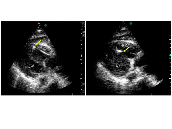
Figure 5.

Use of 2-D echocardiography for monitoring the performance of endomyocardial biopsy in a HT recipient. The arrow indicates the site of the biopsy. Left panel: at the apex of right ventricle, right panel: al the level of the right side of the interventricular septum.
