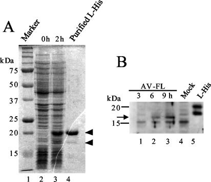
FIG. 3.
(A) Expression of the His-tagged L protein in E. coli and its purification. Lysates of E. coli cells transformed with pET-L were prepared at 0 and 2 h after induction with IPTG and then analyzed by SDS-PAGE. A fraction purified using an Ni affinity column was also analyzed (lane 4). After electrophoresis, the gel was stained with Coomassie blue. The positions of the 20- and 18-kDa proteins are indicated by arrowheads. (B) Synthesis of the L protein in Vero cells transfected with the AV-FL RNA. At the indicated times after transfection by electroporation, cell lysates were prepared and subjected to SDS-PAGE, and then the L protein was detected by Western blotting using antiserum raised against the purified His-tagged L protein. A fraction purified using an Ni affinity column was also analyzed (lane 5). The positions of molecular mass markers are indicated on the left. An arrow indicates the position of the 17-kDa protein.

