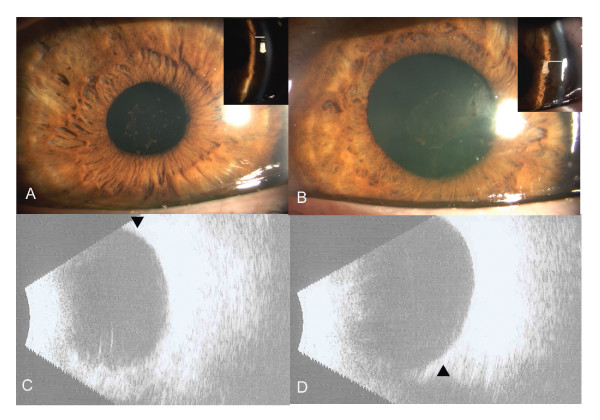
Figure 2.
Slit-lamp photograph at day 5, revealing deep anterior chamber with resolution of corneal edema and conjunctival chemosis in right (A) and Left (B) eyes. Insets: Slit-image showing deep peripheral anterior chamber, depth is marked with line. B) B-scan ultrasound at 2 weeks shows resolution of choroidal effusions (arrow) in Right (C) and left (D) eyes.

