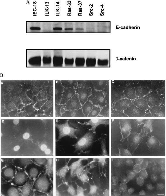
Figure 2.
(A) Immunoblot for E-cadherin and β-catenin. Cell lysates (10 μg) were analyzed for levels of E-cadherin and β-catenin expression by Western blotting as described in Materials and Methods. (B) Indirect immunofluorescence for β-catenin. Cells were plated out on coverslips and stained with antibody toward β-catenin and then with a fluorescent secondary antibody. A) Parental IEC-18; B) control transfected ILK-14 clone A2c3; C) control transfected ILK-14 clone A2c6; D) ILK-overexpressing ILK-13 clone A4a; E) ILK-overexpressing ILK-13 clone Ala3; F) IEC-18GH3IRH kinase deficient ILK; G) IEC-18 cells expressing activated H-ras oncogene (Ras 33) and (Ras 37), respectively; H) Ras 37; I) IEC-18 cells expressing v-src oncogene (Src 2). (Bar = 5 μm.) (×1,000.)

