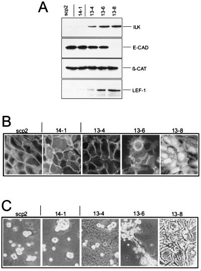
Figure 3.
(A) ILK, E-cadherin, β-catenin, and LEF-1 expression in scp2. Nonidet P-40 cell lysates (10 μg for ILK and LEF-1; 20 μg for E-cadherin and β-catenin) were analyzed by Western blotting as described in Materials and Methods. Clone 14–1: control transfectants, transfected with antisense ILK cDNA; Clones 13–4, 13–6, and 13–8: transfected with ILK-sense cDNA. (B) Indirect immunofluorescence for β-catenin. Cells were fixed in methanol and stained with a mouse mAb for β-catenin (Transduction Laboratories) that was visualized with a fluorescent secondary antibody. (×600.) (C) Morphology on a reconstituted basement membrane gel. Cells were plated on Matrigel, maintained for 72 hr in serum-free medium, and then visualized by phase contrast microscopy. (×150.)

