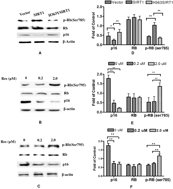
Figure 3. SIRT1-dependend suppression of cellular senescence was through p16INK4A/Rb pathway.
(A) Transfected 2BS cells containing pcDNA3.1, pcDNA-SIRT1 and pcDNA-H363YSIRT1 were lysated and prepared for western blot analysis by using specific antibodies against phospho-retinoblastoma (Ser795), retinoblastoma, p16INK4A and β-actin. (B, C) 2BS cells and WI38 cells at 40 PDs were treated with solvent alone or 0.2 or 2 µM of resveratrol (Res) for 72 h. Cell lysates were prepared for Western blot with the same antibodies. (D, E, F) The quantitative protein expression was summarized in bar graphs (left) presented by mean values (±SEM) of 3 independent experiments. Error bars represent the S.D. Significant difference compared between controls, *, p<0.05; **, p<0.01.

