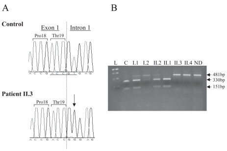
Figure 3.
(A) Electrophoregrams of the normal and mutated GHRHR sequences. Sequence analysis of portion of exon 1 and intron 1 of the GHRHR gene from an unaffected individual (control) and from the proband II.3 (patient). The homozygous T > G transversion (IVS1 + 2 T > G) is indicated by an arrow and the exon 1/intron 1 junction by a dotted vertical line. (B) Segregation analysis of the splice-donor-site mutation of GHRHR in the studied family. A 481-bp GHRHR genomic DNA fragment was digested by Taa-I restriction enzyme that recognizes only the wild type allele (underlined under the electrophoregram), then analyzed on a gel electrophoresis (3% nusieve agarose). This confirmed that the two patients (II.3 and II.4) are homozygous for the mutated allele (481-bp). The parents and the healthy brother (I.1, I.2, and II.2) are heterozygous (three fragments: 418, 330, and 151-bp), the sister (II.1) bears two wild type alleles, and the control (C) has two wild type alleles. L: “1Kb plus” ladder (Invitrogen); ND: not digested, bp: base pair.

