
Figure 3.
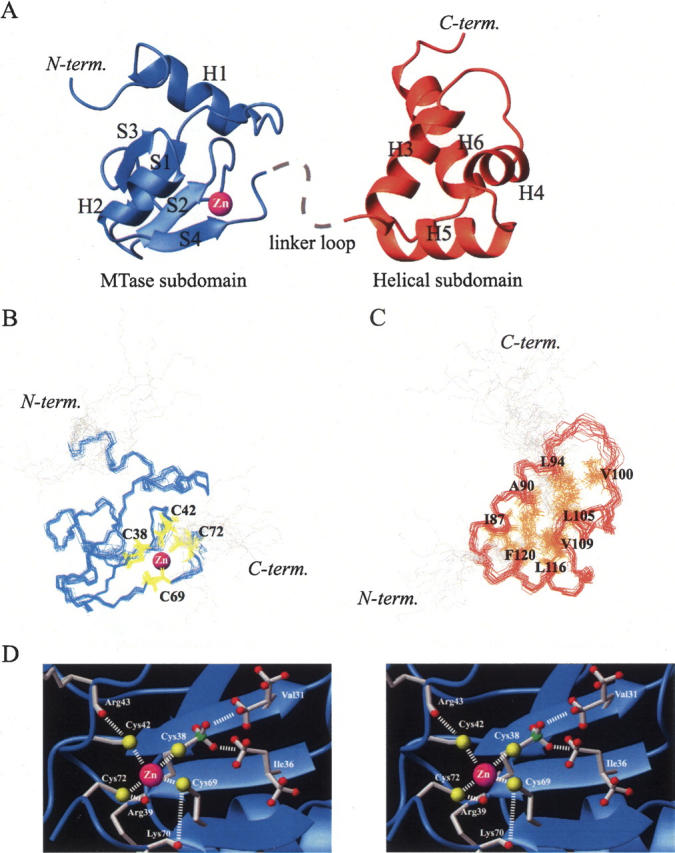
Solution structure of meC38 N-Ada16k. (A) Ribbon representation showing the MTase subdomain (blue) and the helical subdomain (orange). Bound to a protein is the zinc ion (magenta sphere) and the dashed line indicates the linker loop connecting two subdomains. The superposition of 17 lowest energy backbone conformers of (B) the MTase subdomain (blue, residues 9–73) with side chains of Cys38, Cys42, Cys69, and Cys72 (yellow) and (C) the helical subdomain (red, residues 83–123) with side chains of Ile87, Ala90, Leu94, Val100, Leu105, Val109, Leu116, and Phe120 (orange) involved in hydrophobic packing. (D) Zinc–thiolate center of meC38 N-Ada16k in a stereoview. Side chains of Val31, Ile36, Cys38, Arg39, Cys42, Arg43, Cys69, Lys70, and Cys72 are illustrated by stick models. The zinc ion is represented as a magenta sphere. Possible interactions are shown in a dashed line.