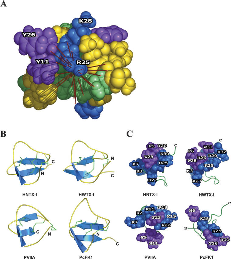
Figure 2.

(A) Putative functional surface of PcFK1 and the dipole moment calculated on each individual NMR solution. The residues are colored green for polar uncharged residues, blue for basic residues, red for acidic residues, purple for aromatic residues, and yellow for aliphatic residues. (B) Secondary structure (blue) and disulfide bridges (green) of Hainantoxin-I (HNTX-I), Huwentoxin-I (HWTX-I), κ-conotoxin PVIIA, and PcFK1. (C) Representation of triplet of residues through which the dipole moment emerges. The residues are colored blue for basic residues and purple for aromatic residues. The figure was done with the PyMOL software.
