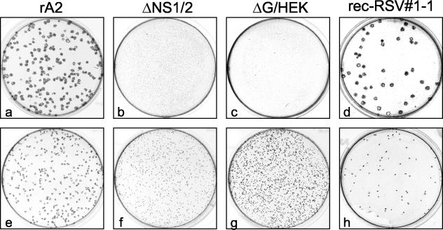
FIG. 3.
Photomicrographs of plaque formation in HEp-2 (a to d) and Vero (e to h) cells infected with rA2 (a and e), ΔNS1/2 (b and f), ΔG/HEK (c and g), or plaque-purified rec-RSV 1-1 (d and h) derived from one of the large plaques identified from the coinfection 1 progeny. Infected cells were incubated for 6 days under methylcellulose, and the plaques were visualized by immunostaining with a cocktail of anti-F monoclonal antibodies.

