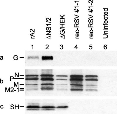
FIG. 8.
Western blot analysis of cell-associated proteins from Vero cells that were infected with either wt rA2 (lane 1), ΔNS1/2 (lane 2), ΔG/HEK (lane 3), plaque-purified rec-RSV 1-1 (lane 4), or plaque-purified rec-RSV 1-2 (lane 5) or were mock infected (lane 6). Western blot analysis was performed using antisera against a peptide representing amino acids 186 to 201 of the G protein (a), purified RSV (b), or a peptide representing amino acids 53 to 64 of the SH protein (c). The relevant cropped portions of blots are shown.

