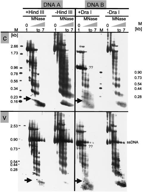
FIG. 7.
Micrococcus nuclease (MNase) digestions revealing the arrangement of nucleosomes in DNA A and B. Nuclei were incubated for different times. Lanes 1, incubation without enzyme for 4 min. Lanes 2 to 7, incubation with enzyme for 0.5, 1, 1.5, 2, 3, or 4 min. One set of aliquots of purified DNAs of every sample was subsequently digested with HindIII (+HindIII) for DNA A or with DraI (+DraI) for DNA B (12 h at 37°C), and the other was left untreated (−HindIII; −DraI). The c-strands (c) and v-strands (v) were hybridized with primer P3a or P4a specific for DNA A or with primer P7c or P8c for DNA B, respectively. The positions of HindIII and DraI sites are indicated in Fig. 4. The sizes of marker bands (M) are shown. Arrows point at the conspicuous swallow-tailed split bands representing mononucleosomes. During the procedure, viral ssDNA was protected by the coat protein. A second prominent band marked with ?? in DNA B might result from subgenomic defective interfering DNA.

