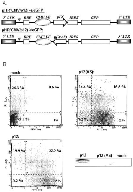
FIG. 1.
p12I expression in Jurkat T cells. (A) Schematic representation of the p12 transgene plasmids pHR′CMV/p12(+)/eGFP and pHR′CMV/p12(−)/eGFP. (B) The HTLV-1 p12I gene was introduced into Jurkat T cells by lentiviral transduction as described in Materials and Methods. Cells were transduced with sense p12I (p12I, expressing both p12I and eGFP) or antisense p12I [p12(AS), expressing eGFP alone] by lentiviral vectors or mock infected. At day 4 posttransduction, an aliquot of cells was stained with propidium iodide (PI) and anti-CD25 antibody by flow cytometry. GFP expression was concurrently analyzed. Approximately 50% of infected cells were found to be GFP positive in both p12I and p12(AS) samples. Samples lysed from approximately 2 million cells were tested for p12I expression by immunoblot assay. The results were representative of three independent infection experiments.

