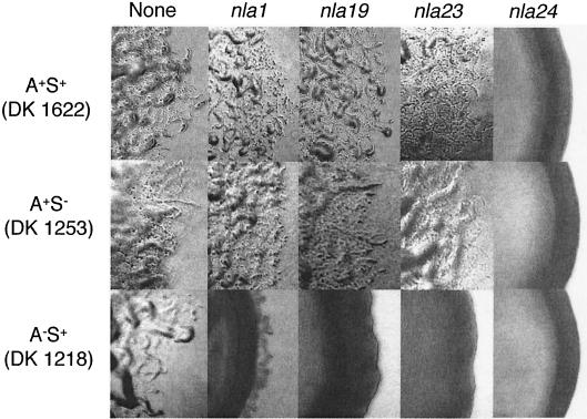
FIG. 4.
Colony edge morphologies produced by nla insertions. The nla1, nla19, nla23, and nla24 insertions were transferred into A+ S+ (DK1622), A− S+ (DK1218), and A+ S− (DK1253) backgrounds, and colony edges were observed using phase-contrast microscopy (×40 magnification). Photographs were taken after 5 days on CTTYE plates.

