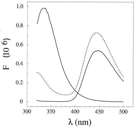
FIG. 3.
Energy transfer from DctD to Mant-ATP. Fluorescence emission was scanned from 320 to 500 nm for DctD (peak emission at 340 nm), Mant-ATP (peak emission at 444 nm), and a mixture of the two (dashed line). Energy transfer from DctD proteins to Mant nucleotide was used to indirectly assay ATP binding. For steady-state data, 290-nm light and 2-nm band pass were used to excite protein (3 μM monomer) or Mant-ATP (100 μM) solutions that were either separate or had been mixed and allowed to set for several minutes at 20°C. Data provided by the manufacturer (PTI, Inc.) were used to correct for variation in the sensitivity of the photomultiplier tube to light of different wavelengths.

