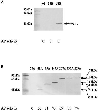
FIG. 7.
Alkaline phosphatase (AP) activities and Western blot analysis of the fusion proteins from the different fusion positions of puc2BA and phoA. The positions of the junction sites are identified by the most C-terminal amino acid residues of Puc2B and Puc2A prior to the PhoA sequence (indicated by arrows in Fig. 6). The cells were grown anaerobically at 100 W/m2 to an OD600 of 0.3 to 0.4. Fifty micrograms of protein from each cell extract was applied to a lane of an SDS-PAGE gel, and anti-PhoA antibody was used to detect the PhoA fusion protein. Assays were performed in triplicate, and the standard deviations were within ± 10%. The values are the averages for the three independent experiments rounded to the nearest integer; a value multiplied by 10−7 is equal to the number of units per cell. The sizes of the hybrid proteins are indicated on the right.

