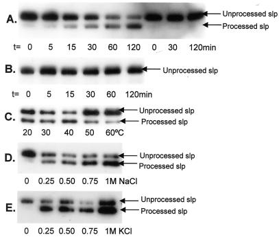
FIG. 2.
Detection of peptidase activity against the truncated S-layer protein. (A) The truncated S-layer protein preparation was used as the substrate in the in vitro peptidase assay with M. voltae membranes as the enzyme source (lanes 1 to 6 from left). Controls with sterile distilled H2O substituting for M. voltae membranes were also performed (lanes 7 to 9 from left). Samples were taken from the reaction mixture at the various indicated time points. Peptidase activity against the His-tagged S-layer protein was detected via Western blotting with anti-His antibodies. (B) The peptidase assay described for panel A was repeated, with heat-treated (95°C for 5 min) M. voltae membranes substituting as the enzyme source. The next three panels show optimization of the signal peptidase assay. The peptidase assays were performed under standard conditions with one changed variable in each case, as indicated. Time courses up to 120 min were performed in each case, but only samples at t = 120 min, at which the reaction appeared to be complete, are shown for comparison. (C) Optimization with respect to temperature. The peptidase assay was performed at 0.4 M KCl with different incubation temperatures as indicated. (D) Optimization with respect to [NaCl]. The peptidase assay was performed at 37°C with different final [NaCl] as indicated. (E) Optimization with respect to [KCl]. The peptidase assay was performed at 37°C with different final [KCl] as indicated.

