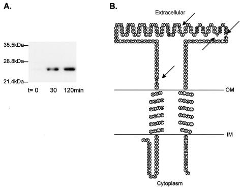
FIG. 3.
(A) Overexpression of the His-tagged M. voltae signal peptidase. The cloned M. voltae signal peptidase was overexpressed in E. coli BL21(DE3)/pLysS. Samples (1.5 ml) were taken preinduction (t = 0) and at 30 and 120 min postinduction and centrifuged, and the pellet was resuspended in 100 μl of ESB and boiled for 5 min. Ten-microliter aliquots were examined by immunoblotting using a primary anti-His antibody at a dilution of 1:20,000. (B) Secondary structure of M. voltae type I signal peptidase, as predicted by TMHMM (17). Arrows indicate the amino acids (Ser-52, His-122, Asp-142, and Asp-148) predicted to be important in activity based on sequence similarity to Sec11. This panel was generated using TOPO transmembrane protein display software (S. J. Johns and R. C. Speth) available online at http://www.sacs.ucsf.edu/TOPO/. OM, outer membrane; IM, inner membrane.

