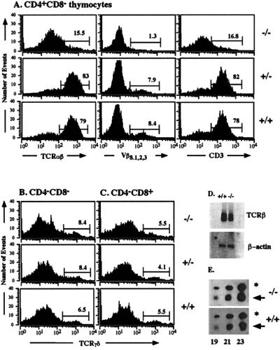
Figure 5.
Normal fetal γδ T cell differentiation but severely reduced numbers of mature CD3high TCRαβhigh CD4+ CD8− SP thymocytes in CREB null mice on E18.5. (A) CD4+ CD8− thymocytes from CREB null mice and control littermates were stained with antibodies against the TCRαβ complex, the TCRVβ8.1,2,3 chain, and CD3ɛ as indicated and were analyzed by flow cytometry. CREB null mice had a severely reduced percentage of mature CD4+ SP cells characterized by high TCRαβ, TCRVβ8.1,2,3, and CD3ɛ surface expression. CD4− CD8− (B) and CD4−CD8+ (C) thymocytes from CREB null mice and control littermates were stained with an antibody against TCRγδ and analyzed by flow cytometry. No difference in γδ T cell differentiation was observed between CREB −/− mice and their control littermates. (D) Northern blot analysis of RNA (500 ng) from sorted DN thymocytes derived from five animals of each genotype. A TCRβ-chain-specific probe containing exon cβ2 was used (Upper). The filter was rehybridized with β-actin as a loading control (Lower). (E) Quantitative RT–PCR using a cDNA derived from sorted DP cells of mutant or wild-type animals, respectively. One representative experiment of five is shown. Gene-specific primers were used for the TCRα chain. Primers for β-actin were used as a standard. Aliquots of the PCR products were taken after 19, 21, and 23 cycles as indicated, separated on an agarose gel, and blotted. Filters were hybridized with internal oligonucleotides to detect the two PCR products: the TCRα product is indicated by an arrow and the β-actin product is indicated by an asterisk.

