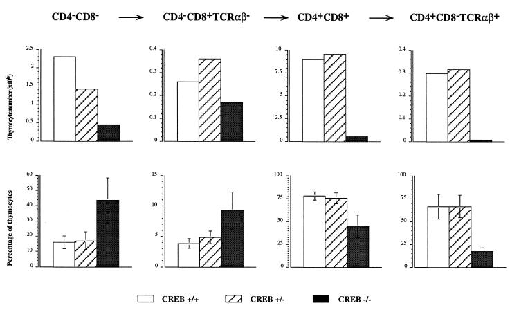
Figure 6.
Altered thymocyte subsets defined by CD4/CD8 surface staining in CREB null mice on E18.5. (Upper) The absolute thymocyte numbers (×106) of each of the thymocyte populations indicated above for one representative animal of each genotype. Open bars, +/+ animal; hatched bars, +/− animal; solid bars, the CREB null mouse. The absolute number of the thymocytes in each of the subsets was decreased in CREB null mice. (Lower) Relative percentage of the thymocyte subsets given as the mean from n = 8 +/+ (open bars), n = 13 +/− (hatched bars), and n = 15 CREB −/− mice (solid bars). The standard deviation is indicated. The relative percentage of CD4− CD8− cells (P < 0.00005, Wilcoxon rank sum test) and immature CD4− CD8+ TCRαβ− SP cells (P < 0.00005, Wilcoxon rank sum test) was increased in CREB null mice, whereas the relative percentage of the more mature CD4+ CD8+ (P < 0.00005, Wilcoxon rank sum test) and the CD4+ CD8− TCRαβ+ SP cells (P < 0.0159, Wilcoxon rank sum test, n = 5) was reduced compared with the littermate controls. We did not observe statistically significant differences between wild-type and heterozygous littermates.

