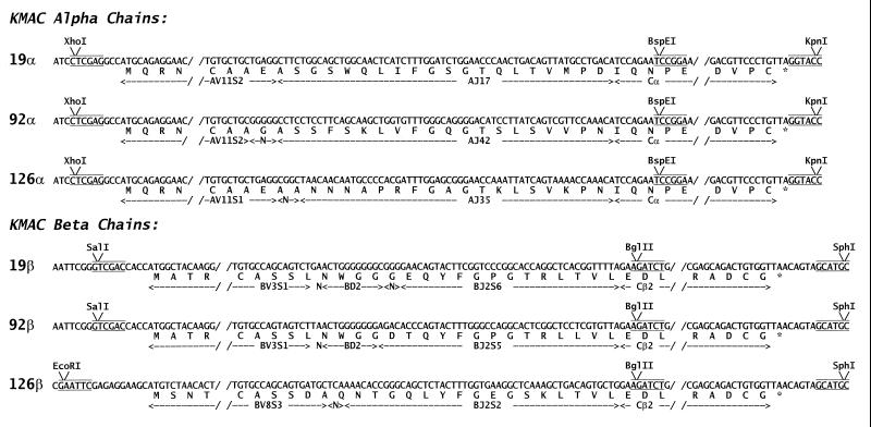
Figure 1.
Sequences of the cloned KMAC TCRs. The figure shows part of the sequences of the KMAC α and β chains as they were cloned into a baculovirus transfer vector. The restriction enzyme sites introduced by the 3′ and 5′ oligonucleotides used to amplify the Vα and Vβ segments are shown. The Cα and Cβ gene segments were truncated as shown just past the codons for cysteines forming the interchain disulfide. In addition the codons for asparagines at position 5 and 118 of Cβ and 80 of Cα were changed to those for aspartic acid to eliminate three N-linked glycosylation sites (not shown).

