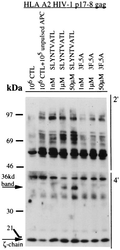
Figure 2.
Protein tyrosine phosphorylation in response to titrations of agonist peptide and antagonist APL. CTL (106) from an HLA A2-restricted line recognizing HIV-1 p17-8 Gag from patient no. 868 were activated by presenting with 105 APC pulsed with either agonist peptide (SLYNTVATL) or the strict antagonist (3F, 5A) at the concentrations indicated above each lane (1 nM, 1 μM, or 50 μM). CTL presented with unpulsed APC were used as a control. Cell lysates were electrophoresed and immunoblotted with anti-phosphotyrosine mAb. For the most detailed depiction, the figure was composed of a 2-min and a 4-min ECL exposure of the same immunoblot.

