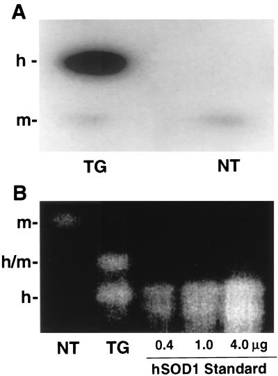
Figure 1.
(A) Expression of SOD1 in TG and NT hearts measured by immunoblotting. Mouse heart tissue was homogenized in buffer containing 25 mM sodium phosphate (pH 7.2), 5 mM EGTA, 1% SDS, and 1 mM phenylmethylsulfonyl fluoride. Total protein (20 μg) was loaded onto a 15% polyacrylamide gel, electrophoresed, and transferred onto nitrocellulose filters. Endogenous mSOD1 and hSOD1 were detected with a polyclonal antibody, pAB-m/h-SOD1, recognizing a common epitope of both mouse and hSOD1. The position of the hSOD1 band is shown by h-, and the position of mSOD1 is shown by m-. (B) SOD1 activity gel of heart tissue extracts (20 μg of protein). The position of the homodimeric mSOD1 is shown by m-, that of homodimeric hSOD1 by h-, and that of the heterodimer by h/m. Both SOD1 protein and activity were increased by approximately 10-fold in the TG hearts compared with that in the NT controls.

