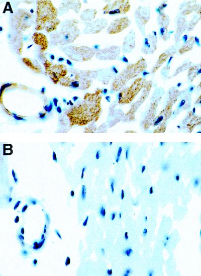
Figure 2.
Immunohistochemical localization of SOD1 in cardiac tissue. Sections were processed for immunocytochemistry (peroxidase-antiperoxidase method) by using the primary antibody to SOD1. The immune reaction was visualized by diaminobenzidine, which results in characteristic brown staining. In the TGs, strong brown staining for SOD1 was seen in both myocytes and endothelial cells, whereas only very weak staining was seen in NT control hearts.

