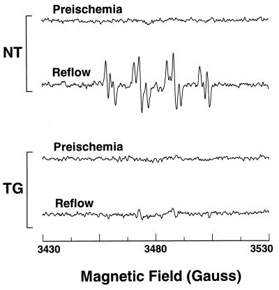
Figure 3.
EPR measurement of free radical generation from TG and NT hearts. Hearts were perfused in the presence of 50 mM DMPO, and effluent was sampled both before ischemia and during the first 2 min of reflow. Measurements were performed at 9.77 GHz with a Bruker ER 300 spectrometer using a TM110 cavity and flat cell with microwave power of 20 mW, and a modulation amplitude of 0.5 G. After reperfusion prominent spectra of DMPO–OOH and DMPO–OH radical adducts were seen in NT hearts but not in TG hearts.

