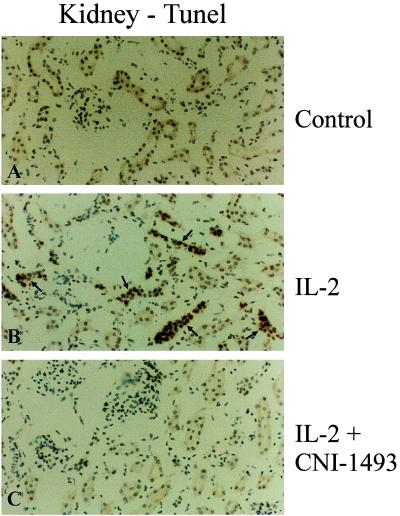
Figure 5.
Immunocytochemical analysis of cellular apoptosis in the kidney by TUNEL staining in untreated control animals (A), IL-2 alone treated animals (B), and IL-2 plus CNI-1493-treated animals (C). Notice the apoptotic cells (arrows) in the sample from an IL-2 alone treated animal (B). Few or no apoptotic profiles were found in comparable sections from controls (A) or the animals treated with IL-2 plus CNI-1493 (C).

