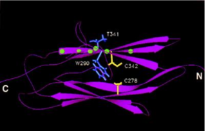
Figure 5.
Molecular modeling of Ig-3 domain of FGFR2. Molecular modeling was used to create a representation of Ig-3 of wild-type FGFR2 based on the crystallographic coordinates of the myosin light chain kinase homolog telokin. A ribbon diagram of the modeled structure is shown indicating the position of the Ig-3 cysteine residues (shown in yellow) relative to the amino acid side chains of W290 and T341 (shown in blue). The mutations W290G and T341P were examined in this study. Balls (shown in green) on the ribbon diagram indicate the positions of other noncysteine craniosynostosis mutations in the Ig-3 domain (Table 1C).

