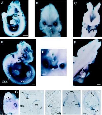
Figure 6.
Spatial distribution of Arp1 transcripts during mouse development. (A) Lateral view of E9.5 embryo. Hybridizable RNA is observed in the abdominal area (Abd), in surface ectoderm of the maxillary (Mx), and mandibular (Ma) regions of the first branchial arch. Arrowhead points out the mesenchymal component of the mandibular arch. Staining of the optic vesicle (Ov) is because of the nonspecific trapping of colored conjugates as evidenced by reaction with a sense probe. No signal was observed in spinal cord (Sc). (B) Anterior view of the head of E9.5 embryo. Facial part has been removed for better visualization of the signal. Staining is observed in the developing Rathke’s pouch (Rp) and in two bilateral structures (marked by white asterisks), still to be identified. (C) Anterior view of the first branchial arches of E9.5 embryo. Arrowheads point to the sharp bands of Arp1 expression along the surface ectoderm of the mandibular component of the first branchial arch. (D) Lateral view of E10.5 embryo. Expression is detected in a number of new structures: in the dermomyotomal compartments of the somites (dmy), in the presumable migratory myogenic precursors (of the limb muscle) localized in the ventrolateral myotome (vlmy) at the forelimb level. The signal is also seen at the periphery of the eye. Strong staining is observed in the restricted domains of the forebrain (white arrows) at the area of the cranial flexure (cf). Black triangle corresponds to the midbrain/forebrain boundary; fl, forelimb; hl, hindlimb. (E) High magnification of the anterior view of head of E10.5 embryo (facial part has been removed); expression of Arp1 is sharply localized to the Rathke’s pouch. Bilateral staining previously detected becomes stronger and more compact. (F) Dorsolateral view of E10.5 embryo. Arp1 specific signal is seen in presumable ventral (v) and dorsal (d) myoblast cells within the forelimb (fl). (G) Anteriolateral view of E10.5 head showing bilateral distribution of Arp1 signal in the forebrain. (H) Sagittal section of the prestained E10.5 embryo. Expression is stronger in the pars dislis (PD) than in pars intermedia (PI) within the Rathke’s pouch. Expression is seen in the ectoderm and mesenchyme (arrowhead) of the mandibular branch (Ma) of the first branchial arch. (I) Staining was detected on sagittal section in epithelial cells of the bronchi (arrow) within the lung bud (Lb). (J and K) Transverse sections of the E10.5 embryo at the level of the forelimbs. Open triangle points to the myoblast precursor cells migrating into the proximal portion of the forelimb. Staining is also revealed in the mesenchyme of the dorsal mesentery (Dm); G, gut. [Bars = 200 mm (A, D, and F–I), 100 mm (J and K), and 50 mm (B and C).]

