
Fig. 6.
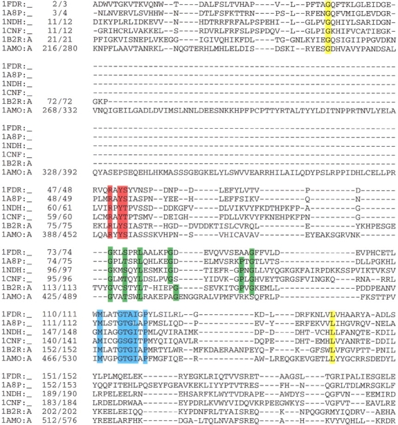
Sequence alignment of all FAD-containing proteins resembling the ferredoxin reductase structure based on structure-sequence alignment as obtained using the CE program (Shindyalov and Bourne 1998). Conserved sequence motifs are depicted in boxes: RxYS(T) in red, GxxS(T)xxL(x)5G(x)7PxG in green, and MxxxGT(S)G(A)IxP in blue. In yellow are conserved residues not interacting with the cofactor. The data bank accession code and position numbers according to the sequence and according to the PDB are given. Proteins: 1FDR, flavodoxin reductase from Escherichia coli; 1A8P, ferredoxin oxidoreductase from Azotobacter vinelandii; 1NDH, cytochrome b5 reductase from Sus scrofa; 2CND, nitrate reductase from Zea mays; 1AMO, NADPH-cytochrome P450 reductase from Rattus norvegicus; 1CQX, flavohemoprotein from Alcaligenes eutrophus.