
Fig. 7.
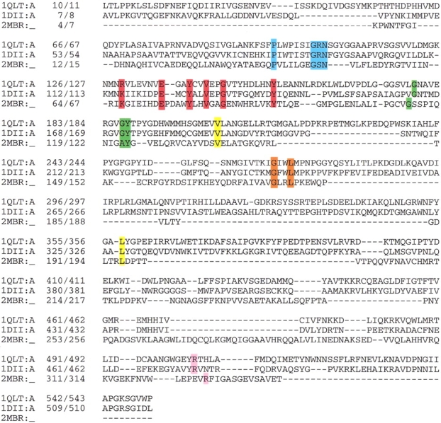
The sequence alignment of FAD-containing proteins resembles the p-cresol methylhydroxylase structure. Conserved sequence motifs are depicted in boxes: R(x)6ExxYxxVxxG(x)8Y in red, P(x)6G(A)xN in blue, GxxL in orange, R in pink, and G(x)8GY in green. The data bank accession code and position numbers according to the sequence and according to the PDB are given. Proteins: 1DII, p-cresol methylhydroxylase from Pseudomonas putida; 1QLT, vanillyl-alcohol oxidase from Penicillium simplicissimum; 2MBR, uridine diphospho-N-acetylenolpyruvylglucosamine reductase from Escherichia coli.