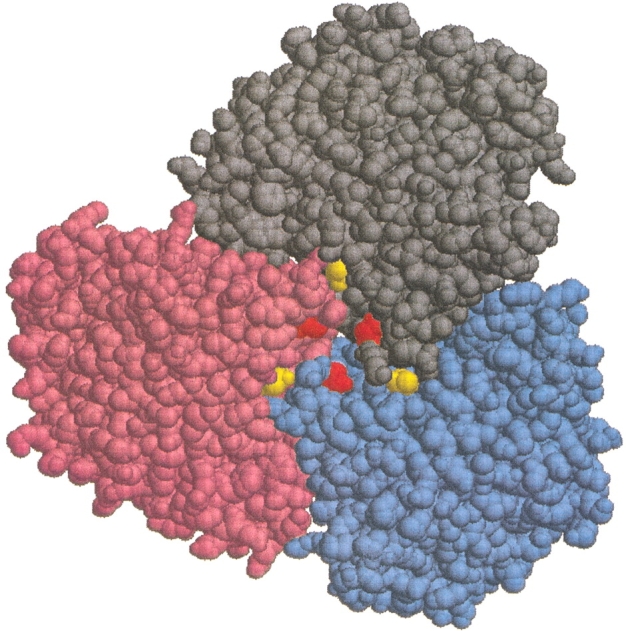
Fig. 1.

Thermococcus litoralis wild-type glutamate dehydrogenase viewed along the threefold (trimer-trimer) axis with the top trimer removed, thus exposing the trimer-trimer interface. Subunits A, B, and C are colored gray, blue, and pink, respectively, and the Thr138s and Asp167s are colored yellow and red, respectively. The distance between any two Asp167s is ∼10 Å, while that between any two Thr138s is ∼20 Å. Figure created using the program RasMol.
