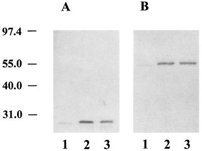
Figure 2.
Western blot analysis of cAb 26–2F. Reduced proteins (400 ng) were separated by SDS/10% PAGE and transferred to nitrocellulose sheets. These were incubated with either goat anti-human κ chain (A) or goat anti-human IgG Fc-specific (B) antibodies followed by treatment with alkaline phosphatase-labeled rabbit anti-goat IgG and nitroblue tetrazolium. Lane 1, mAb 26–2F; lane 2, cAb 26–2F from S13–1; lane 3, cAb 26–2F from P4–5. Molecular weight standards (×10−3) are at left.

