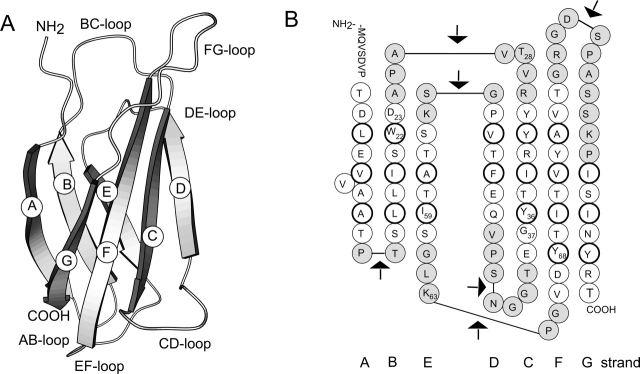
Figure 1.
(A) Schematic drawing of the structure of FNfn10 (Dickinson et al. 1994). (B) The amino acid sequence of FNfn10 in its secondary structure context. Residues in a β-strand are shown as white circles. Those residues whose side chain forms the hydrophobic core are enclosed with a thicker line. Loop residues are shown shaded. The arrows mark the position in the loops where FNfn10 was separated to generate complementary fragments.

