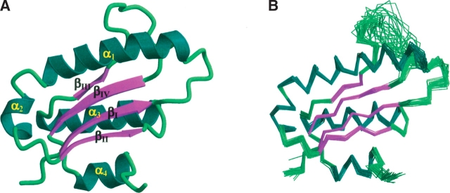
Figure 1.
(A) Ribbon diagram of the NMR structure of AF2095 for residues 1–112 colored by secondary structure. Disordered residues 113–123 were removed for clarity. (B) Superposition of the backbone (N,C,C′) atoms for the 30 best structures determined for AF2095 for residues 1–112. The disorder for residues Q79–I91 in the loop that connects β-strands β3 and β4 is evident by the large RMSD spread. The figures were generated with MOLSCRIPT (Kraulis 1991) and rendered with Raster3D (Merritt and Bacon 1997).

