
Figure 2.
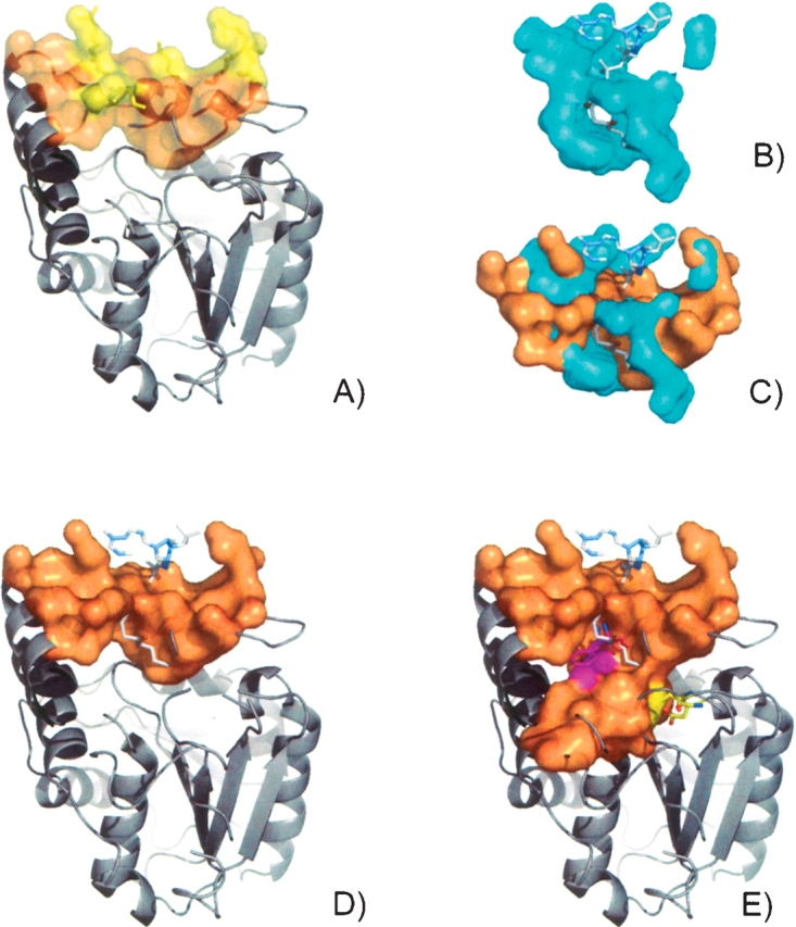
The surface pocket (CASTp ID 20, orange) on BioH from E. coli (PDB ID 1m33) contains a grouping of basic residues (yellow) (A). The CoA binding from aminoglycoside 2′-N-acetyltransferase (AAC(2′)-lc) from M. tuberculosis (PDB ID 1m4g, E.C.2.3.1.-) (B). CoA ligand has been modeled into the surface of BioH (D), based on the superposition with AAC(2′)-lc (C). Using a smaller solvent probe radius (1.2 Å) to define the pocket, the surface reveals an additional channel protruding into the domain interface which contains the buried catalytic triad (yellow) and Phe143 (magenta) (E).