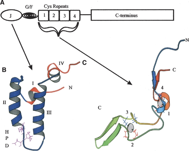
Figure 1.
Structural motifs found in E. coli DnaJ. (A) A diagrammatic representation of the domains present in E. coli DnaJ. J represents the J domain, G/F stands for the Gly/Phe-rich region, and cysteine repeats represents the four zinc-finger-like motifs. The first three domains correspond to approximately half DnaJ. (B) A ribbon representation of the J domain (1XBL) (Pellechia et al. 1996). The conserved HPD motif is depicted in purple. The four helices are labeled. (C) A ribbon representation of the cysteine repeats (1EXK) (Martinez-Yamout et al. 2000). Cysteine residues are depicted in red (repeats 1 and 2) and blue (repeats 3 and 4). Repeats 1 and 4 form zinc center 1 and repeats 2 and 3 form zinc center 2 (Linke et al. 2003). The coordinated zinc atoms are shown in CPK format. This diagram is not to scale. The figures were generated using Molscript (Kraulis 1991).

