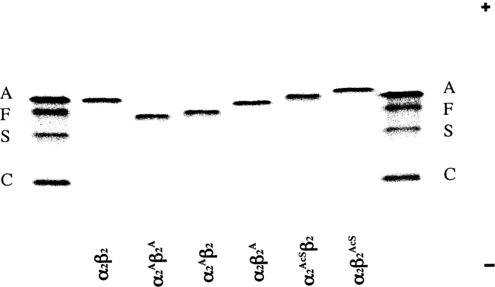
Figure 2.
Isoelectric focusing of purified recombinant Hb mutants. Approximately 5 μg of each Hb was applied to a pH 6.0–8.0 range gel (Hb Resolve, Perkin-Elmer Life Sciences). Electrophoresis was performed for 30 min at 600 V and then for 45 min at 900 V at 10°C. The gel was stained with the JBZ stain (Perkin-Elmer-Wallach). The anode is at the top and the cathode is at the bottom. The standard hemoglobins on the far left and the far right are hemoglobins A, F, S, and C. The identity of each recombinant hemoglobin is shown under each lane. A small superscript “A” indicates an N-terminal Ala on that subunit. A small superscript “AcS” indicates an N-terminal acetylserine on that subunit. Subunits without superscripts contain N-terminal Val, e.g., α2β2 is HbA.

