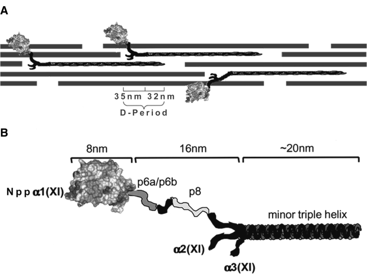
Figure 1.
Schematic of collagen fibril and Npp α1(XI) collagen. (A) Collagen fibril shown comprised of collagen types II, IX, and XI. The globular domain Nppα1(XI) collagen is shown to extend from the surface of the collagen fibril. The dimensions of the gap-overlap region of the D-period are indicated. (B) Schematic representation of the Npp α1(XI) and adjacent variable region and minor triple helix. The dimensions as determined (Gregory et al. 2000) by transmission electron microscopy are indicated. The position of the Npp α1(XI) domain is indicated as distal with respect to the adjacent p6a/p6b/p8 variable region and minor triple helix. The α2(XI) and α3(XI) chains are also indicated in the diagram.

