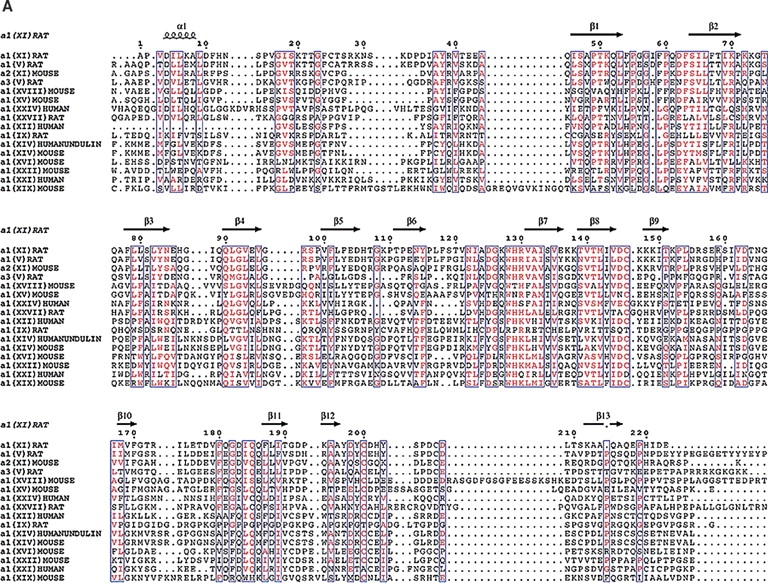
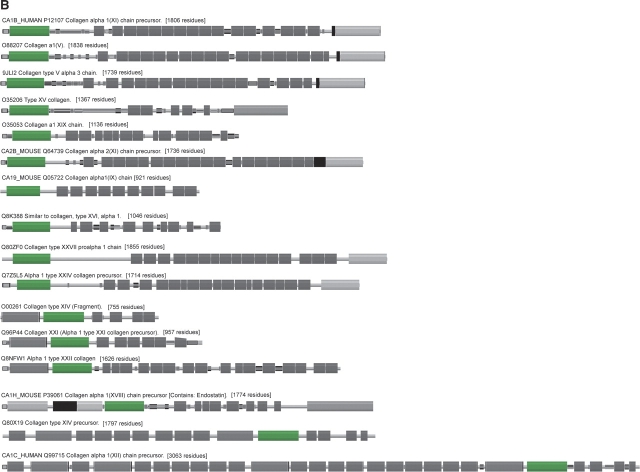
Figure 9.

A comparison of collagens containing similar LNS domains. (A) Identical residues are red while similar residues are purple. The β-strands fall in highly conserved regions suggesting that our model may extended to other collagens that contain the LNS or thrombospondin-like domain. The β-strand which contains the putative heparin binding site is unique to Npp α1(XI) collagen and not conserved among the other collagens. The highly conserved cysteine residues suggest that they may be important in the structure of this domain. (B) Pfam schematic of LNS (TSPN) domain (shown as green box) in collagens. This domain is contained in a number of collagens but may vary in position.

