Figure 3.
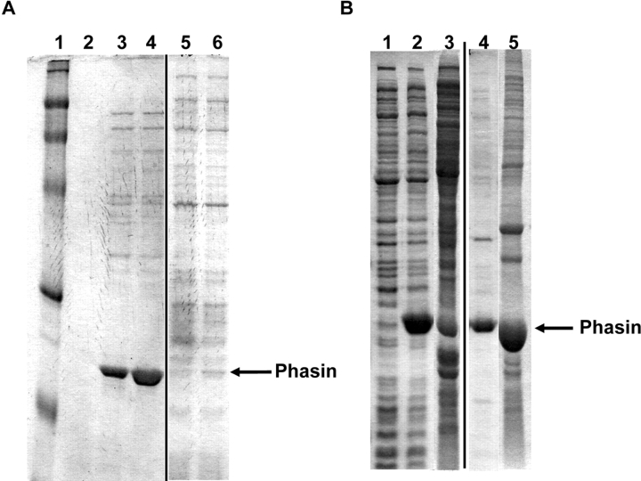
SDS-PAGE results for phasin affinity to PHB. (A) BLR strain carrying phaP gene (plasmid pET/phaP) induced for 0.5 and 2 h at 37°C. Lane 1, molecular weight marker. Lane 2, preinduction whole-cell lysate. Lanes 3 and 4, soluble fractions of cell lysates at 0.5- and 2-h inductions, respectively. Lanes 5 and 6, insoluble fractions corresponding to lanes 3 and 4. (B) BLR strain carrying the phaP gene (plasmid pET/phaP) and PHB biosynthesis genes (plasmid pJM9131) grown and induced for 8 and 30 h. Lane 1, preinduction whole-cell lysate. Lanes 2 and 3, soluble fractions after 8 and 30 h, respectively. Lanes 4 and 5, insoluble fractions corresponding to lanes 2 and 3. Note the displacement of phasin from the soluble fraction (B, lane 2) to the insoluble fraction (B, lane 5) in the presence of PHB (after 30 h of growth).

