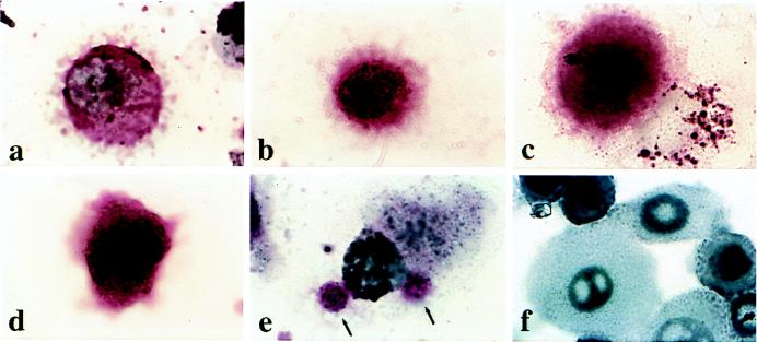
Figure 3.
Expression of cytokeratin and Muc-1 glycoprotein in circulating breast and prostate carcinoma cells and normal epithelial cells. Circulating tumor cells (a-e) were isolated by using the ferrofluid purification followed by cytospinning and staining of the slide. (a) A cell stained with anti-mucin-1 from a patient with metastatic breast cancer. (b) Same patient but a different cell stained with anti-cytokeratins 5, 6, 8, and 18. (c) A cell stained with anti-cytokeratin from a patient with clinically organ-confined breast tumor. (d) A cell stained with anti-cytokeratin from a patient with clinically organ-confined prostate cancer. (e) Same patient as in a showing two stained bodies, probably apoptotic tumor cell bodies (arrows), stained with anti-cytokeratin and attached to a macrophage. (f) Normal epithelium obtained from human trypinized foreskin (uncultured) and stained with anti-mucin-1. All slides were subjected to an alkaline-phosphatase-anti-alkaline-phosphatase procedure that caused the development of a red precipitate. The nuclei were counterstained with hematoxylin. All images were photographed at ×1,000.

