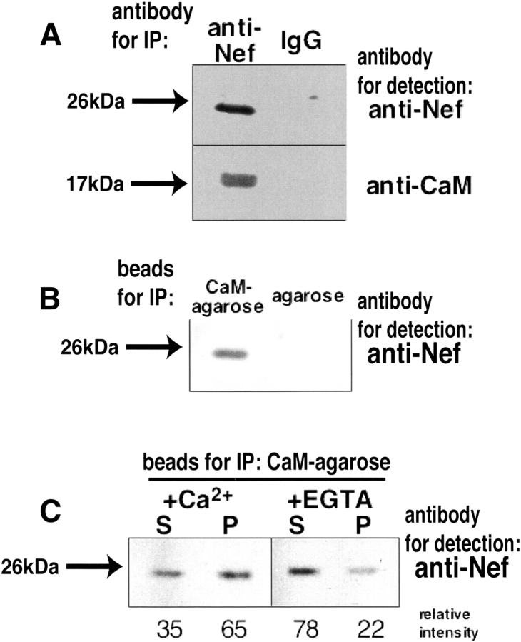
Figure 1.
Immunoprecipitation analyses of Nef expressed in NIH3T3 cells. (A) Immunoblot analysis of Nef and calmodulin coimmunoprecipitated from lysates of NIH3T3 cells, which were permanently infected with Nef expression vector. Cell lysates were immunoprecipitated (IP) with the anti-Nef antibody (left lane) and a normal IgG used as a negative control (right lane), and were detected by anti-Nef (upper) and anti-CaM (lower) antibody, respectively. (B) NIH3T3 cells permanently expressing Nef were lysed and incubated with calmodulin-agarose beads (left) or agarose beads without calmodulin (right) in the presence of 2 mM CaCl2. After washing of the agarose beads, the bound proteins were resolved by SDS-PAGE and transferred to membrane. Blots were probed for anti-Nef antibody. (C) Cells were lysed, centrifuged to be separated into soluble and insoluble fraction, and incubated with calmodulin-agarose beads in the presence (left) or absence (right) of CaCl2. S and P represent the soluble fraction and the insoluble fraction, respectively. Blots were probed for anti-Nef antibody. Relative intensities of the bands are also shown under the gel image.

