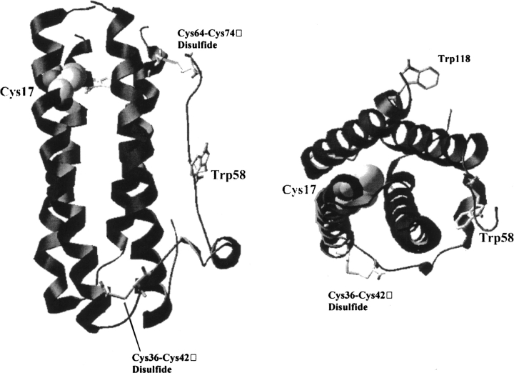
Figure 1.
Crystal structure of recombinant human G-CSF. Shown are ribbon diagrams of G-CSF. The diagram on the left provides a view orthogonal to the long axis of the protein. On the right, the diagram is rotated 90° to give a view down the axis of the 4-helix bundle. The two native disulfides (36–42 and 64–74) and tryptophans 58 and 118 are shown as stick representations. Cysteine 17, the only free thiol in the protein, is shown as a space-filling model. The disulfide bond 64–74 was removed from the image on the right for clarity.

