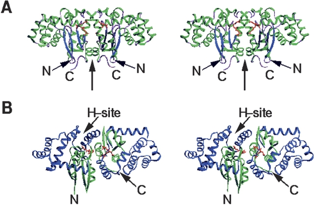
Figure 1.
Overall structure of the dimeric hGSTK. Two GSF molecules are shown as red ball-and-stick models. (A) View perpendicular to the twofold NCS axis (thick arrow), showing the butterfly-like shape of the dimer. The α-helices and β-sheets are shown in green and in blue, respectively. (B) View showing the binding cleft of the H-site. Domains I and II are shown in green and in blue, respectively. These diagrams were prepared using the program SETOR (Evans 1993).

