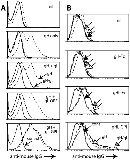
Figure 1. Expression of gH/gL as a single Fc fusion protein.
A. 293T cells were transfected with different expression plasmids. 48h later they were trypsinized and analyzed by flow cytometry for expression of gH with mAb 8C1 (short dashes) or gH/gL with mAb 7E5 (long dashes). Control = secondary antibody only (solid lines). “gH+gL ORF” used the full-length genomic ORF47; “gH+gL” used the RACE-mapped gL, which starts at the 5th in-frame ORF47 AUG codon [9]; “gH+gL-GPI” used a GPI-linked form of the RACE-mapped gL. In each case, gH was expressed from the full-length genomic ORF22. nil = untransfected. B. 293T cells were transfected with diferent gH expression plasmids or left untransfected (nil), and 48h later analyzed for gH expression with mAb 8C1 (short dashes, white arrow) and for gH/gL expresion with mAb 7E5 (long dashes, grey arrow), as in A. Solid lines/black arrow = secondary antibody only. gHL-GPI and gHL-Fc are the same fusion protein with either a C-terminal GPI anchor or human IgG1-Fc. Each histogram shows 10,000 cells. Both gH-specific and gH/gL-specific staining was significantly increased after gHL-Fc transfection compared with controls (p<0.00001 by Student's t test). Equivalent results were obtained in a repeat experiment.

