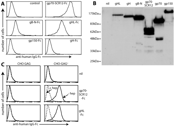
Figure 2. gHL-Fc binds to GAGs.
A. Supernatants from 293T cells transfected with expression constructs for different IgG-Fc fusion proteins were used to stain BHK-21 cells. gp70-SCR12 = short consensus repeats 1 and 2 of gp70, which include its heparin-binding site; gB-N = gB N-terminal to its furin cleavage site, which incorporates its cell-binding domain. To retain GAG expression, the cells were plated onto Petri dishes 18h before staining and then detached without trypsinization. Control = untransfected 293T cell supernatant plus secondary antibody. Equivalent data were obtained in 4 repeat experiments. B. The transfected cell supernatants from A were immunoblotted for their common human IgG-Fc domain. nil = untransfected cell supernatant. gp70 = Fc fusion of the full-length protein, which was not used here. C. GAG+ CHO cells or the GAG-deficient CHO-745 mutant (GAG−) were stained with Fc fusion proteins as indicated, either in the presence (dashed lines) or absence (solid lines) of 300 µg/ml heparin. nil = secondary antibody only. Equivalent data were obtained in 2 repeat experiments.

