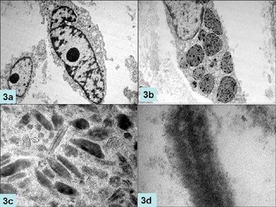
Figure 3.

Electronmicrographs showing oval to spindle shaped cells with prominent nucleoli (a, ×2250 original magnification) packed with compound melanosomes and premelanosomes (b, ×6250); higher magnification of premelanosomes demonstrating internal structure (c, ×8250) and electron dense neurosecretory granules (d, ×6250).
