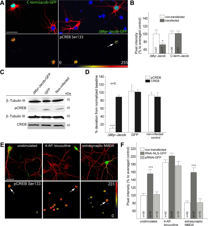
Figure 9. Nuclear Jacob Regulates the Phosphorylation of CREB.
Primary hippocampal neurons were transfected at DIV11, fixed the next day, and stained with MAP2-specific antibodies (Cy5), pCREB (Ser133; Alexa568), and mounted with DAPI-containing (blue) medium. Gradient lookup tables applied to determine the dynamic range of pCREB are included to visualize pixel intensity differences as indicated with the scale from 0 to 255. Scale bar indicates 40 μm.
(A and B) Overexpression of ΔMyr-Jacob-GFP decreases the basal level of CREB phosphorylation in nonstimulated primary hippocampal cultures (DIV12). No effect as compared to nontransfected control neurons was seen after the transfection of a Jacob-GFP construct encoding the C-terminal half of Jacob. Quantification is shown in (B). A single asterisk (*) indicates p < 0.01.
(C and D) Overexpression of ΔMyr-Jacob-EGFP using a Semliki Forest virus vector significantly reduced the level of pCREB at resting conditions in comparison to EGFP-infected and noninfected cortical primary neurons. The diagram (D) represents the data from four to five independent experiments normalized to β-Tubulin III. Total CREB levels were not affected by infection of the cultures.
(E and F) Knockdown of nuclear Jacob utilizing the construct RNAi-NLS-GFP increases basal pCREB levels and prevents CREB shut-off after stimulation of extrasynaptic NMDA receptors. Prior to bath application of NMDA, cultures were stimulated with bicuculline in the presence of MK801. Arrows indicate RNAi-NLS-GFP–transfected neurons. Cultures were fixed 30 min after the stimulation with either bicuculline or the subsequent bath application with NMDA. Triple asterisks (***) indicate p < 0.001.

