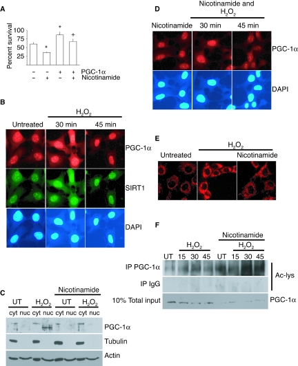
Fig. 2.
SIRT1 regulates PGC-1α subcellular localization and activity in response to stress. (A) Percent survival of cells after exposure to H2O2 (350 µm, 1 h) with or without nicotinamide (10 mm); values represent means ± standard error of the mean; * indicates significant difference in survival compared to control cells; + indicates significant difference in survival compared to peroxide-treated control cells (P < 0.05). (B) Immunofluorescent detection of PGC-1α and SIRT1 in cells following treatment with H2O2 (350 µm). Nuclei were visualized with DAPI stain. (C) PGC-1α in cytoplasmic (cyt) and nuclear (nuc) subcellular fractions in cultured cells after treatment with H2O2 (350 µm, 45 min), with or without nicotinamide (10 mm). (D) Immunofluorescent detection of PGC-1α in nicotinamide-treated cells (10 mm) after treatment with hydrogen peroxide (350 µm, 45 min). (E) Mitotracker Red detection of mitochondrial membrane potential after exposure to H2O2 (350 µm, 1 h) in nicotinamide- (10 mm) treated cells. (F) Detection of acetylated PGC-1α by Western in immunoprecipitates from cells following exposure to hydrogen peroxide (350 mm), with and without nicotinamide (10 mm).

