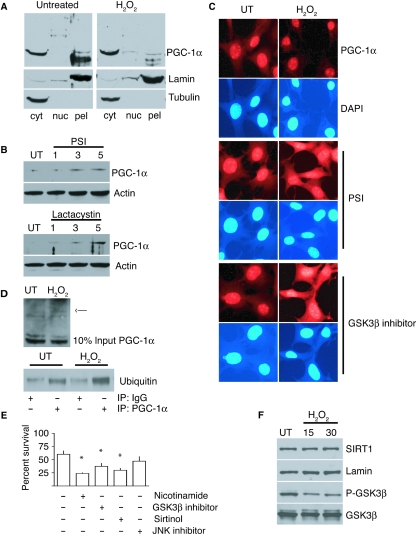
Fig. 3.
PGC-1α is targeted for GSK3β-dependent proteasomal degradation in response to oxidative stress. (A) Detection of PGC-1α protein by Western blot in subcellular fractions from cells 1 h after treatment with hydrogen peroxide (350 µm). (B) Detection of PGC-1α protein by Western blot in whole cell lysates from cells treated with proteasomal inhibitors PSI (10 µm) and lactacystin (10 µm) for the indicated times in hours. (C) Immunofluorescent detection of PGC-1α after treatment with hydrogen peroxide (350 µm, 45 min); cells were grown under normal conditions or with proteasomal (PSI 10 µm) or GSK3β (GSK3β inhibitor VIII 20 µm) inhibitors prior to and during stress. (D) Detection of ubiquitinated species by Western blot of PGC-1α immunoprecipitates from untreated and hydrogen-peroxide-treated cells (350 µm, 45 min). (E) Percent survival of cells after exposure to hydrogen peroxide (350 µm, 1 h); cells were grown under normal conditions or pre-incubated with nicotinamide (10 µm), GSK3β Inhibitor VII (20 µm), sirtinol (25 µm) or c-Jun N-terminal kinase (JNK) inhibitor II (25 µm); values represent means ± standard error of the mean; * indicates significant difference in survival compared to peroxide-treated control cells (P < 0.05). (F) Western blot detection of SIRT1, phospho-GSK3β and GSK3β in extracts taken at the times indicated in minutes from cells exposed to hydrogen peroxide (350 µm).

